
Tabouri Sarah1*, Bentchouk Jesia Asma2, Belkralladi Houria3, Khaldi Houria4, Maaref Nawel5, Bouhenni Hakima5 and Merazga Djamel Eddine6
1Department of Medical Oncology, Center for the Fight Against Cancer of Sidi Bel Abbes, Taleb Morad Faculty of Medicine – Djillali Liabes University, Algeria
2Breast and Pelvic Surgery Department – Center for The Fight Against Cancer of Sidi Bel Abbes, Taleb Morad Faculty of Medicine – Djillali Liabes University, Algeria 3Department of Pathology – University Hospital HASSANI Abdelkader – Taleb Morad Faculty of Medicine – Djillali Liabes University, Algeria
4Department of Radiotherapy-Oncology – Center for the Fight Against Cancer of Sidi Bel Abbes, Taleb Morad Faculty of Medicine – Djillali Liabes University, Algeria 5Department of Medical Oncology, Center for the fight against cancer of Sidi Bel Abbes, Algeria
6Department of General Surgery – Mascara Public Hospital Establishment, Algeria
*Corresponding Author: Tabouri Sarah, Department of Medical Oncology, Center for the Fight Against Cancer of Sidi Bel Abbes, Taleb Morad Faculty of Medicine – Djillali Liabes University, Algeria
Abstract
The frequency of breast tissue outside the mammary gland, or supernumerary breast, is rare, with a reported incidence in women of 0.3 to 6% [1]. This tissue is typically located in the armpit [1,2], ectopic tissue can be the site of benign or malignant physiological variations similar to normally located breast tissue. The axilla is the most common site, while the sternal, infra-clavicular, epigastric, and vulvar regions have also been described.
We report a case of infiltrating lobular carcinoma of the axillary supernumerary breast managed at the Tidjini HADDAM Cancer
Center in Sidi bel abbes in ALGERIA.
Keywords: Supernumerary Breast; Axillary Breast; Infiltrating Lobular Carcinoma; Breast Cancer
Introduction
Extra-mammary or supernumerary breast tissue is rare, with a reported incidence of 0.3 to 6% [1]. Breast embryonic develop- ment begins during the 4th week of gestation and is located on the ventral surface of the body. The «milk line» forms a ridge from the bilateral mammary ectodermal tissue and extends from the base of the axillary region to the inguinal region.
From the 5th week of gestation, the «milk line» gradually disap- pears but continues to proliferate and differentiate into mamma- ry lobules specifically at the level of the 4th intercostal space [2]. Failure and/or abnormality of this involution process can lead to ectopic breast tissue presenting as polymastia (accessory breast glands) or polythelia (accessory nipples) [3].
Ectopic breast tissue has the same anatomical and physiologi- cal characteristics as breast tissue in the «normal» position and is subject to the same hormonal repercussions, vulnerable to thesame disorders, including malignant transformation [4].
Indeed, ectopic breast cancer accounts for 0.2 to 0.6% of all breast cancers [5]. The axilla is the most common location (70- 90%), with the glandular tissue located in the subcutaneous tissue and deep dermis [6,7]. Its diagnosis is mostly delayed, estimated at
40.5 months [8] due to misdiagnoses, especially with other subcu- taneous masses: lymphadenopathy, sebaceous cysts, nevi, lipomas, or hidradenitis [4].
Case Report
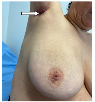
We report the case of a 61-year-old female patient who present- ed to our clinic with an unlabeled axillary retraction for 12 years, but who recently presented with unusual paroxysmal axillary pain and a change in coloration with the appearance of pseudo-pustular erythema without any breast signs. Clinically, the patient had a red, 5 cm major axis, axillary cutaneous mass, adherent to the cutane- ous and deep planes, polylobulated, with palpable, round, 3 cm diameter adenopathies fixed to the deep plane. The homolateral breast is normal in appearance, with congenital nipple umbilica- tion which is also bilateral (Figure 1).
An initial axillary ultrasound was performed and revealed a he- terogeneous anechoic axillary cutaneous mass with deep extension measuring 38x17mm, in close contact with the axillary vascular axes; associated with a 34x24mm axillary lymph node mass.
A breast MRI confirmed the presence of a neoplastic process on a supernumerary breast and ruled out any breast lesions in both breasts.
A microbiopsy revealed breast parenchyma with diffuse car- cinomatous proliferation arranged in «Indian files», composed of medium to large round cells with marked cytological atypia and high mitotic index. This appearance corresponds to a grade II inva- sive lobular carcinoma according to SBR, on supernumerary breast. Immunohistochemistry confirmed an invasive lobular carcinoma of the Luminal B type with 80% positive hormone receptors, HER2 negative and Ki67 at 10%.
After an extension assessment, namely a thoraco-abdomi- no-pelvic scan and bone scintigraphy, the tumor was classified as T4dN2M0, and neoadjuvant chemotherapy followed by surgery and external radiotherapy was decided after a multidisciplinary consultation meeting.
The patient was placed on neoadjuvant chemotherapy, protocol 04 AC 60 – 12 Paclitaxel, i.e. 04 cycles combining adriamycin and cyclophosphamide followed by 12 cycles of paclitaxel at 80mg/m². A minor clinical response was observed after the 04 AC60 cycles, but the redness disappeared and the mass became mobile relative to the deep plane after the 12 paclitaxel cycles (Figure 2).

The patient underwent surgery, which consisted of a lenticular resection of the entire tumor mass and a lymph node dissection, without any procedure on the homolateral breast.
The histological and immunohistochemical study on the surgi- cal specimen found at gross description a tissue formation mea- suring 10/07/03 cm covered by a skin. slice of section: presence of a well-defined whitish nodule of 1.5 cm.
Microscopic description found a Tumor cells arranged in sheets, single files and large nests.Tumor cells are discohesive, small, mo- nomorphic with scant cytoplasm and low grade nuclei. The tumor stroma is abundantly fibrous and inflammatory. Peri-neural inva- sion is present. lymph node dissection found 10 lymph nodes infil- trated by the same neoplastic process out of the 19. The histologi- cal response was classified as RCB1 (Residual Cancer Burden1) and the tumor was classified as pT1c N3a (Figure 3).
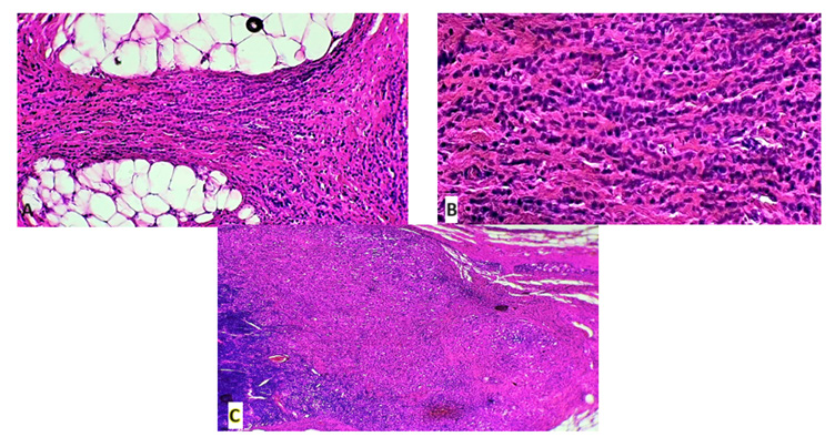
Immunohistochemistry: Estrogen and progesterone receptors was positive, respectively 70% and 80%, KI-67 (60%) and HER2 score 2 with FISH test negative.
The disease progression was marked on the 17th postoperative day by the onset of homolateral mastitis of the lower quadrants with significant inflammatory signs, paroxysmal pain, and a fever of 39°C without signs of parietal sepsis. We requested a breast MRI, which confirmed the homolateral mastitis with extensive dermo- hypodermic cellulitis of the entire breast and post-operative fibro- scarring without signs of malignancy. The patient responded to a 10-day antibiotic treatment with Pristinamycin and Metronidazole with complete clinical and radiological response.
The patient received external beam radiation therapy to the ax- illary region at a dose of 45Gy.
Discussion
Polymastia is a congenital condition characterized by the per- sistence of breast tissue in an abnormal location. It is located along the primitive mammary line, which extends from the armpit to the inguinal fold. It is estimated that 0.3% to 6% of women and 1% to 3% of men have a supernumerary breast. There are two types of ectopic breast tissue: supernumerary breast tissue and aberrant breast tissue [9,10]. Supernumerary breasts include a nipple, areo- la, and mammary gland, either isolated or associated [11].
Teir reported that the frequency varied between different eth- nic groups, ranging from 0.6% to 6% [11,12]. Regarding aberrant breast tissue, they have neither nipple nor areola, and they gener- ally do not involve a secretory system [11]. They are usually locat- ed near the breasts [13]. The armpit is the most common location, while the sternum, infra-clavicular region, epigastrium, and vulva have also been described [9,11,14]. Evans., et al. found ectopic breast tissue in the armpit in 71% of cases [4] and in 58% of cases for Marshall., et al. followed by the sternal region in 18.5%, sub clavicular in 8.6%, sub mammary in 8.6%, and vulvar in 4% [11]. Overall, they are associated with a frequency that varies between 1.7% and 6%. In 13% of cases, an axillary supernumerary breast is present on both sides [15].
Like normal breast tissue, ectopic breast tissue is hormone-de- pendent. The anomaly is present at birth but is usually only discov- ered during pregnancy or lactation, when it may increase in size, become cyclically painful, or cause discomfort. Suspected abnor- mal ectopic breast tissue follows the same diagnostic principles as normal tissue. This includes anamnesis, physical examination, and conventional paraclinical tests [16]. It is important to note that the presence of a supernumerary breast may go unnoticed on standard mammograms due to its location. The tumor may be located in a place that does not appear on standard radiographic images. Ul- trasound should be used to complement mammography. MRI may be performed to exclude ipsilateral or contralateral breast cancer.
Since ectopic breast tissue can be bilateral, contralateral evaluation should be strongly considered [7]. In our case, an MRI was recom- mended and performed due to the lobular histological type of the carcinoma [17,18]. The reported incidence of the lobular histologi- cal subtype among ectopic breast carcinomas is 3 to 9.5% [1,19]. A review of the Japanese literature showed that medullary, muci- nous, and apocrine carcinomas were more common. Additionally, Marshall., et al. noted that the distribution of histological types was as follows: 79% invasive ductal carcinomas, 9.5% invasive lobular carcinomas, and 9.5% medullary carcinomas [11].
Histologically, the absence of lymph node tissue and the pres- ence of normal breast ducts and lobules adjacent to the tumor are necessary to confirm the diagnosis of ectopic breast carcinoma and to exclude the possibility of a metastatic tumor from an occult pri- mary lesion [20]. Ectopic breast cancer is classified using the same TNM staging system as for primary breast cancer. Due to its sub- cutaneous location, skin involvement may be early and lead to T4 stage cancer [19]. In half of the cases, there is extension of the can- cer to the axillary lymph nodes at the time of diagnosis. This early nodal extension is explained by the proximity of the malignancy to the nodes, but also by the delay in diagnosis, which can be 3 to 24 months after the onset of symptoms, especially in the absence of a nipple and areola.
The diagnosis and management of ectopic breast cancer follow the same therapeutic approach as breast cancer. Due to its rarity, there is no acceptable level of evidence to support a different ap- proach. A high index of suspicion is necessary to diagnose carci- noma of an ectopic breast. Once the diagnosis is established, it is important to assess the extent of tumor spread, which includes a bone scan, chest-abdominal CT scan, and breast MRI to ensure the absence of distant metastases and a second breast cancer.
Due to its incidence and the scarcity of data, surgical manage- ment has been controversial in recent years. Historically, ectopic breast cancer has been treated with radical mastectomy and chest irradiation [19]. Later, Cogswell and Czerny [15] demonstrated that ipsilateral mastectomy in these patients does not lead to a bet- ter prognosis. In a series of autopsied patients with ectopic breast cancer who died of metastatic lesions in the skeletal system, pleu- ra, and liver, there was no infiltration of the ipsilateral breast. No breast involvement was observed in radical mastectomy specimens in patients with distant metastases. Evans and Guyton [7] conclud- ed that there was no additional benefit of mastectomy compared to wide excision with axillary lymph node dissection in the presence of sentinel node positivity. These aggressive techniques are no lon- ger an option, and local excision is the recommended procedure. Mastectomy should only be considered in patients with a second primary breast lesion.
Regarding adjuvant treatment, it has the same indications as breast carcinoma in its anatomical position [20]. This treatment
can be supplemented with hormone therapy when the tumor is positive for hormone receptors (positive estrogen receptors).
It is difficult to accurately assess the prognosis of breast cancer on ectopic breast tissue because the diagnosis is often delayed and because of the lack of long-term follow-up data for this rare tumor.
A Japanese retrospective study of 98 patients reported in the journal Surgical Oncology that, of 64 evaluable patients, all but one were alive after a median follow-up of 28 months [21]. A 2015 Chi- nese retrospective study published in Oncology Letters included 11 cases of breast cancer on ectopic breast tissue [22]. The patients, aged 27 to 48 years, were followed for a median of 20 months. Axil- lary lymph node dissection was performed in six of them. Four had distant metastases. The three-year overall survival rate was 54.5% (6 of 11 patients) due to the occurrence of late metastases. In 2011, Strasbourg surgeons reported in the journal Gynecology Obstetric and Fertility a case of cancer on ectopic breast tissue that occurred several years after its surgical removal. No cases of cancer after ex- cision of an accessory breast gland had been previously published. The tumor had developed on the scar of the ectopic breast gland excision.
Breast cancer on ectopic breast tissue can also occur in male patients. In a literature review published in 2011 in the Journal of Plastic, Reconstructive and Aesthetic Surgery, Dutch and Ital- ian surgeons reported six cases in men among 171 cases of axil- lary breast carcinomas (with an average delay of 40 months before diagnosis) [8] and its incidence on ectopic breast tissue is higher than in the usual form of male breast cancer [24].
Conclusion
Ectopic or supernumerary breast carcinoma is a rare and diffi- cult diagnosis due to the lack of information and awareness among clinicians.
Furthermore, supernumerary axillary breast tissue is not de- tected by most screening and diagnostic tests. Its management is based on a multidisciplinary approach following the same recom- mendations as for breast cancer. It can also occur in men and does not seem to have a worse prognosis when diagnosed at similar stages of the disease.
Bibliography
1. Schmidt H. “Supernumerary nipples: prevalence, size, sex and side predilection – a prospective clinical study”. European Jour- nal of Pediatrics 157 (1998): 821-823.
2. Georgiade NG., et al. “The Breast: Embryology, Anatomyand Physiology in Aesthetic Surgery of the Breast 1st ed”. WB Saun- ders, Philadelphia (1990).
3. Bland KI and Romrell LJ. “Congenital and Acquired Distur- bances of Breast Development and Growth. The Breast. Com- prehensive Management of Benign and Malignant Diseases”. Bland KI, Copeland EM 3rd (ed): WB Saunders, Philadelphia (1991).
4. Velanovich V. “Ectopic breast tissue, supernumerary breasts, and supernumerary nipples”. South Medical Journal 88 (1995): 903-906.
5. Francone E., et al. “Ectopic breast cancer: case report and re- view of the literature”. Aesthetic Plastic Surgery 37 (2013): 746-749.
6. Nihon-Yanagi Y., et al. “A case of ectopic breast cancer with a literature review”. Surgery on Oncology 20 (2011): 35-42.
7. Evans DM and Guyton DP. “Carcinoma of the axillary breast”. Journal of Surgical Oncology 59 (1995): 190-195.
8. Visconti G., et al. “Approach and management of primary ecto- pic breast carcinoma in the axilla. “where are we? A compre- hensive historical literature review”. Journal of Plastic, Recon- structive and Aesthetic Surgery 64 (2011): E1-11.
9. Amsler E., et al. “Ectopic breast cancer of the axilla”. In Annales de dermatologie et de venereologie 129.12 (2002): 1389-1391.
10. Copeland MM and Geschickter CF. “Diagnosis and treatment of premalignant lesions of the breast”. Surgical Clinics of North America 30.6 (1950): 1717-1741.
11. Marshall MB., et al. “Ectopic breast cancer: case report and lit- erature review”. Surgical Oncology 3.5 (1994): 295-304.
12. Aubert JP., et al. “Glande mammaire surnuméraire: un cas in- habituel”. In Annales de chirurgie plastique et esthétique. 36.5 (1991): 457-459.
13. Haddad H., et al. “Cancer du sein sur tissu mammaire ec- topique: à propos de 02”. Pan African Medical Journal 13 (2012): 50.
14. Routiot T., et al. “Breast carcinoma located in ectopic breast tissue: a case report and review of the literature”. Oncology Re- port 5.2 (1998): 413-420.
15. Cogswell HD and Czerny EW. “Carcinoma of aberrant breast of the axilla”. American Surgery 27 (1961): 388.
16. Khan RN., et al. “Invasive carcinoma in accessory axillary breast tissue: a case report”. International Journal of Surgery Case Reports 59 (2019): 152-155.
17. Salemis NS. “Primary ectopic breast carcinoma in the axilla: a rare presentation and review of the literature”. Breast Disease 40.2 (2021): 109-114.
1. Selvi V., et al. “Role of magnetic resonance imaging in the pre- operative staging and work-up of patients affected by inva- sive lobular carcinoma or invasive ductolobular carcinoma”. BioMed Research International 2018 (2018): 1-8.
2. Algara M., et al. “Radiation techniques used in patients with breast cancer: results of a survey in Spain”. Reports of Practical Oncology and Radiotherapy 17 (2012): 122-128.
3. Roorda AK., et al. “Ectopic breast cancer. “special treatment considerations in the postmenopausal patient”. Breast Journal 8.5 (2002): 286-289.
4. Matsuoka H., et al. “A case of carcinoma originating from ac- cessory breast tissue of the axilla. (Article in Japanese)”. Gan No Rinsho 30 (1984): 387-391.
5. Zhang S., et al. “Diagnosis and treatment of accessory breast cancer in 11 patients”. Oncology Letter 10 (2015): 1783-1788.
6. Guèye M., et al. “Cancer du sein sur la cicatrice d’éxerese d’un tissu mammaire ectopique: à propos d’un cas”. Gynécologie Ob- stétrique and Fertilité 39.3 (2011): e55-57.
7. Yamamura J., et al. “Male breast cancer originating in an acces- sory mammary gland in the axilla: a case report”. Case Reports on Medicine 2012 (2012): 286210.