
1Department of Medical Oncology, Center for the fight against cancer of Sidi Bel Abbes. Taleb Morad Faculty of Medicine – Djillali Liabes University
2Department of Gynecology and Obstetrics, Mother and Child Hospital of Sidi Bel Abbes. Taleb Morad Faculty of Medicine – Djillali Liabes University
3Department of Pathology and Cytology – Sidi Bel Abbes University Hospital Center. Taleb Morad Faculty of Medicine – Djillali Liabes University
4Breast Surgery Department – Center for the fight against cancer of Sidi Bel Abbes. Taleb Morad Faculty of Medicine – Djillali Liabes University
5Independent Pathologist – Sidi Bel Abbes
Abstract
Breast angiosarcomas are rare mesenchymal tumors of vascular endothelial origin, most commonly associated with radiation, and less frequently primary. They are characterized by a distinct clinical and radiological presentation, frequent local recurrence, and a poor prognosis. We report the case of a young patient who presented with primary angiosarcoma in both breasts, occurring a year apart. Subsequently, she developed bilateral ovarian metastases, treated with surgery and radiotherapy for breast tumors, and underwent palliative chemotherapy for ovarian metastases. Diagnostic challenges were encountered in both radiological and pathological aspects. Through this case, we discuss the clinical, radiological, and pathological features of asynchronous bilateral primary angiosarcoma of the breast with ovarian metastases.
Keywords: Angiosarcoma, Breast, Metastases, Ovaries
Introduction
Breast sarcomas are extremely rare malignant tumors, accounting for less than 1% of breast tumors and less than 5% of all sarcomas; their incidence is estimated to be less than 50 cases per 10 million women [1]. Breast angiosarcomas represent 0.04 to 1% of all malignant breast tumors [2-3]. They occur either spontaneously in women under 40 years old, referred to as « primary, » or after thoracic radiotherapy in older patients [4]. Definitive diagnosis is histological, requiring additional immunohistochemical studies to confirm the vascular nature of angiosarcomas, expressing endothelial markers such as CD31 and CD43 [5]. Surgery is the primary treatment [6]. We present the case of a 32-year-old patient who developed asynchronous bilateral angiosarcoma in both breasts, followed by metastatic relapse in the ovaries within a very short period. Through this case, we will discuss the epidemiological, diagnostic, and therapeutic aspects of these rare tumors.
Case Presentation
This is 32-year-old patient, nulligeste, with no specific medical history, who initially consulted for a swelling in the left breast that had been evolving for a year. Clinical examination revealed a nodule at the junction of the inner quadrants, measuring 6 cm in diameter, painless, mobile in relation to the deep plane and the skin, without orange peel texture or inflammatory signs. Mammography identified a very low-density opacity with indistinct borders, measuring
66.8mm in the major axis, located in the inner quadrants of the left breast; additional ultrasound of the breasts revealed a heterogeneous echogenic structure with indistinct borders in the inner quadrants. The mass was classified as ACR 3, and the axillary lymph node areas were free.
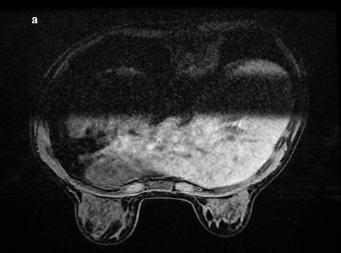
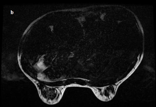
A microbiopsy of the nodule was performed, and histological analysis revealed a benign mesenchymal proliferation, concluding it to be an Angiolipoma. The patient underwent surgery three months later, with a tumorectomy performed. Histological examination again confirmed an Angiolipoma with insufficient excision margins. Six months later, the patient returned for another consultation due to a rapidly enlarging local recurrence. Clinical examination revealed a 5 cm mass in the inner quadrants. Breast MRI was conducted, revealing a left breast mass in the inner quadrants measuring 64 mm, showing hyperintensity in STIR in T2, hypointensity in T1, with gradual centripetal enhancement mainly at its lower pole, revealing fatty areas without signs of infiltration of the pectoral muscle. This mass was vascular, suggesting either an Angiolipoma or angioma, and was classified as ACR 3 (Figure 1).
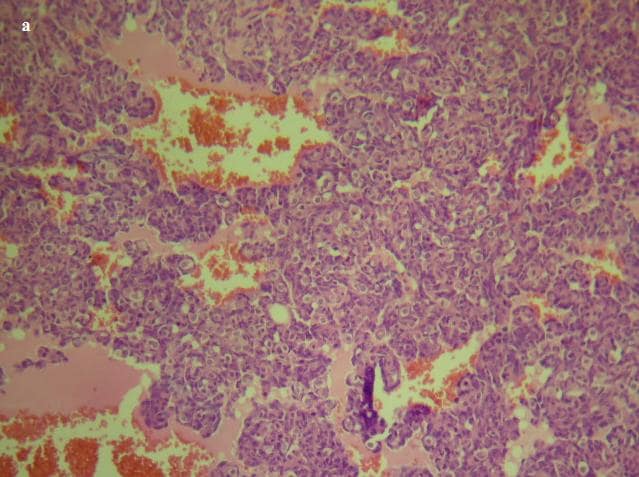
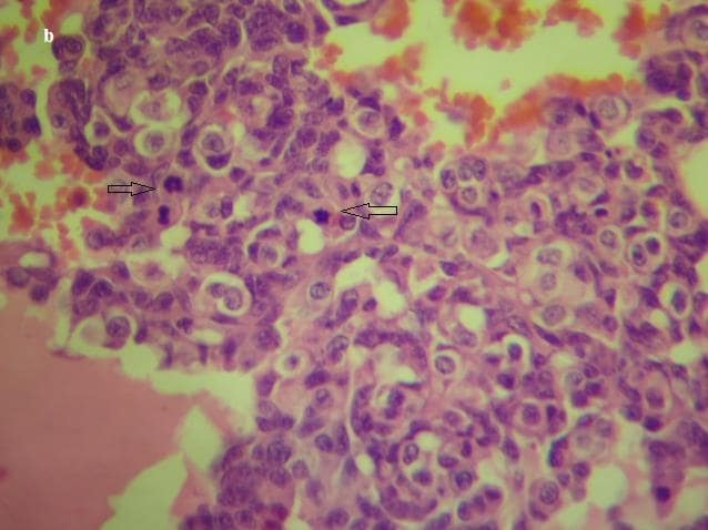
The patient underwent a wide local excision (zonectomy), and the histopathological study confirmed the diagnosis of intermediate-grade angiosarcoma of the breast (Figure 2).
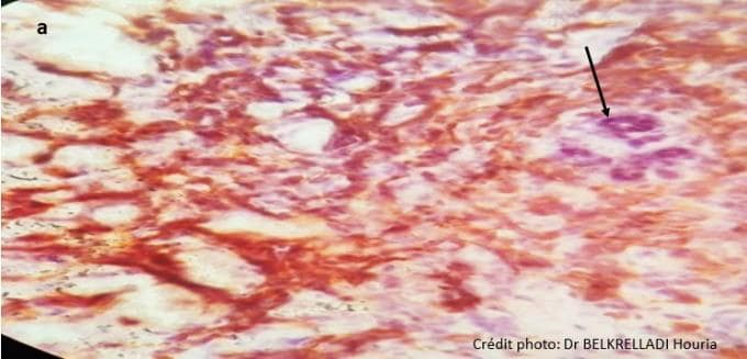
An immunohistochemical study was performed to confirm the diagnosis, revealing positivity of the tumor cells for CD31 and D34 (Figure 3).
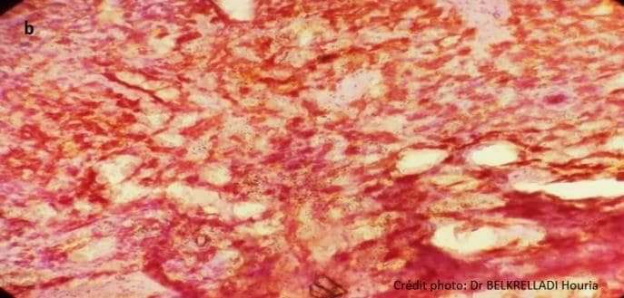
After six months, the patient experienced a local recurrence and underwent reoperation, this time undergoing mastectomy. Histopathological analysis revealed an intermediate-grade angiosarcoma of the breast with clear surgical margins, located 1.5mm from the deep plane. The patient received adjuvant radiotherapy, specifically 3D conformal radiotherapy, at a dose of 50 Gray with a conventional fractionation schedule, 2 Gray per session, 5 sessions per week. The target volume was the thoracic wall with 3mm 3D margins.
Four months after the completion of radiotherapy, the patient presented with a newly appearing mass in the right breast. Breast MRI identified a 22mm mass classified as ACR3, suggesting either an angiolipoma or angioma. However, a biopsy confirmed the malignant nature of the tumor. The patient underwent surgery, undergoing total mastectomy, followed by radiotherapy using the same protocol as for the left breast tumor.
Three months after the completion of radiotherapy, the patient sought consultation due to a desire for pregnancy. However, during pelvic ultrasound, a right lateral-uterine mass measuring 20cm was discovered. An MRI was
requested, revealing two large bilateral ovarian masses with morphological characteristics inconsistent with their dynamic features. The dynamic features were more indicative of ovarian hyperplasia, contrasting with a morphological appearance suggestive of a secondary involvement. The radiologist concluded that the diagnosis favored secondary involvement.
An exploratory laparotomy was decided upon, and intraoperatively, hypervascularized nodular masses were found bilaterally in the ovaries. On the right side, the mass was excised with preservation of the ovarian parenchyma, while on the left side, where the mass involved the entire left ovary, a left salpingo-oophorectomy was performed.
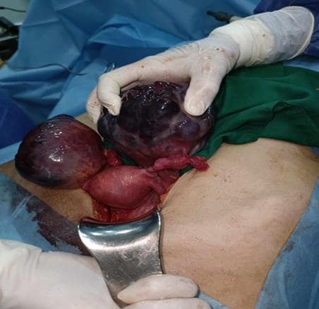
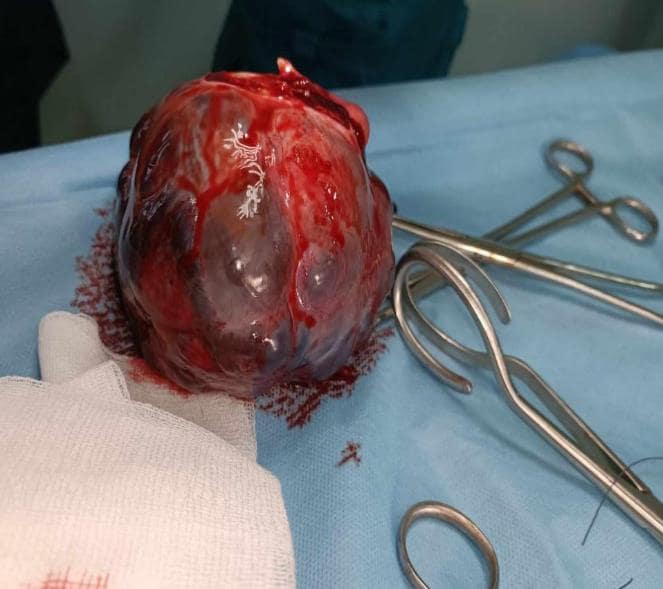
The histopathological examination of the operative specimen reveals a pattern of infiltrating mesenchymal cellular proliferation, mostly composed of solid areas associated with vascular structures of variable sizes, sometimes anastomosed. The tumor cells are round, ovoid, spindle-shaped, with indistinct borders, eosinophilic cytoplasm, and irregular vesicular nucleoli. Cytonuclear atypia and mitoses are moderate. The tumor stroma is delicate with extensive areas of necrosis and hemorrhage. The left tubal wall shows congestive hemorrhagic changes without signs of tumor infiltration. Cytological analysis of the ascitic fluid reveals a clear background traversed by polymorphous inflammatory elements mixed with clusters and sheets of tumor cells.
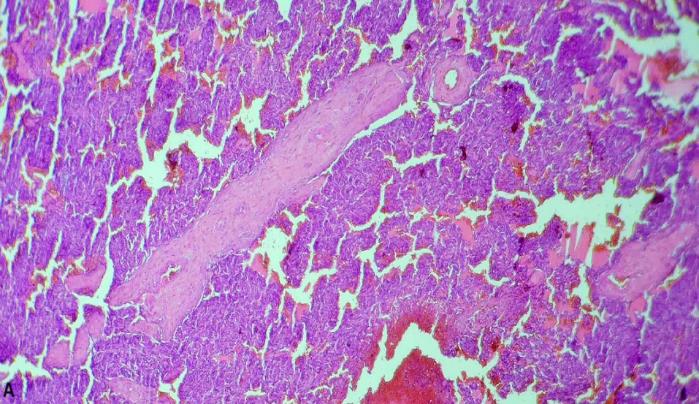
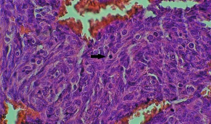
Immunostaining was performed (Figure 6) revealing positive nuclear staining with almost diffuse distribution for ERG antibody on tumor cells, and negative staining for Pancytokeratin.
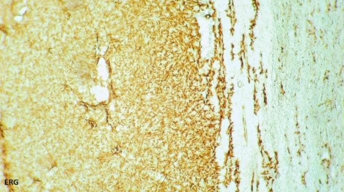
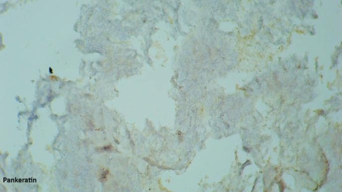
The diagnosis was thus established as the histopathological and immunohistochemical features of bilateral ovarian metastases from a high-grade angiosarcoma without signs of infiltration of the left fallopian tube, along with ascitic fluid containing malignant tumor cells.
The postoperative extension assessment, including a thoraco- abdominal CT scan and pelvic MRI, revealed no anomalies except for residual right ovarian parenchyma. Following a multidisciplinary discussion, palliative chemotherapy with Paclitaxel was decided for the patient.
Discussion
Breast sarcomas are rare malignant tumors, encompassing phyllodes tumors of grade I and II, as well as other entities forming a group that includes grade III phyllodes tumors and angiosarcomas. The 3-year relapse-free survival for patients with grade I and II phyllodes tumors is 57%, 45% for grade III phyllodes tumors, and 7% for angiosarcomas [6].
Angiosarcomas represent 0.04% of all malignant breast tumors and 8 to 10% of breast sarcomas. They typically occur in women aged between 30 and 50 years, but can affect all age groups, ranging from 13 to 85 years [3].
Although the pathogenesis is unknown in the majority of angiosarcoma cases, several etiological factors, such as a history of irradiation, chronic lymphedema, and genetic syndromes like Recklinghausen syndrome, Maffucci syndrome, and Xeroderma pigmentosum, are established as playing a significant role in this type of sarcoma. Approximately 3% of primary angiosarcomas are associated with a genetic alteration [7].
Angiosarcomas can manifest either as tumors in young women that occur spontaneously without an apparent trigger or as tumors in older women (average age of 69 years) arising after radiation therapy as part of breast cancer conserving treatment [8]. These radio-induced sarcomas occur 5 to 10
years (median at 6 years) after exposure to 50 Grays of radiation.
Bilateral breast angiosarcomas are even rarer, with only a few cases reported in the literature, including one case in a breastfeeding woman [9].
Clinically, the typical presentation is the rapid enlargement of a breast nodule, as observed in our patient. Tumor size is often substantial, ranging from 2 to 11 cm, with an average of
5.3 cm [9]. The tumor may also be revealed by the sudden onset of a hematoma. Two signs suggestive of breast angiosarcoma are the pulsatile nature of the tumor, found in some cases, and the purplish angioma-like appearance of the skin overlying the tumor. These characteristics were absent in our patient.
Mammographic appearance is nonspecific and can be misleading, mimicking a benign lesion, as was the case in our patient. The tumor may appear as a rounded or lobulated opacity, without calcifications, low density, homogeneous, often well-circumscribed, and may show focal density asymmetry [10]. In young women with dense breasts, the tumor is often challenging to visualize on mammography. On ultrasound, breast angiosarcoma appears as a tissue mass (hyperechoic, hypoechoic), heterogeneous with areas of fluid indicative of hemorrhagic phenomena [11]. It can also present as a mixed echostructure (both hyperechoic and hypoechoic) without a true mass. Color Doppler analysis shows intense hypervascularity [10].
MRI reveals a heterogeneous mass with low T1 signal and high T2 signal, sometimes with areas of spontaneous T1 hyperintensity corresponding to hemorrhagic areas or venous lakes [11]. MRI also helps assess subcutaneous infiltration and visualize large draining vessels [12]. This examination plays a crucial role in the assessment of local extension (especially infiltration of deep muscle planes). It is noteworthy that cases of secondary angiosarcomas following radiotherapy are characterized by foci within thickened, irradiated skin.
Histology is the only examination that allows for a definitive diagnosis. The term « angiosarcoma » encompasses all malignant tumors whose cells exhibit morphological and functional properties of normal endothelium. Macroscopically, angiosarcomas are invasive tumors, poorly demarcated, with hemorrhagic areas. Microscopically, their differentiation is highly variable.
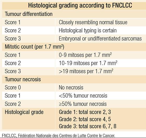
Distinguishing them from atypical post-radiation vascular lesions (in the context of a history of radiotherapy) or even benign lesions (hemangioma or angiolipoma) can be challenging in some cases. In the case of our patient, two histological readings concluded it was a benign tumor, which did not align with the disease’s progression.Some authors report that the histological grade of the FNCLCC for soft tissue sarcomas (Table 1) is applicable to primary breast sarcomas [12]. However, this grade is not routinely applied for primary breast angiosarcomas. Instead, a histological classification specific to breast angiosarcomas is used, namely the classification proposed by Donnell, et al. [13].
Immunohistochemistry is essential to confirm the vascular nature of the lesion. Angiosarcomas typically express endothelial markers: CD31 (the most sensitive and specific marker), CD34, factor VIII, Ulex europaeus agglutinin 1, and vascular endothelial growth factor (VEGF) [14].
The cornerstone of the treatment for breast angiosarcomas is surgery, which remains the only curative approach. Similar to other sarcomas, it involves a wide excision with R0 resection. This surgical intervention may entail either conservative treatment or radical mastectomy. Simple lumpectomy may be considered for small tumors less than 3cm [15]. Salvage mastectomy remains an option in cases of local recurrence after conservative treatment, particularly for small tumors less than 5 cm and of low grade [16]. The adequacy of surgical margins influences the likelihood of local recurrence. Axillary lymph node dissection is not recommended due to the low probability of lymph node involvement in these tumors [17,18].
Angiosarcomas are highly aggressive tumors, and as a result, 50% of patients will develop local recurrences or distant metastases [19].
Adjuvant chemotherapy is controversial and does not appear to provide a benefit; thus, it is not recommended. However, there may be a potential benefit in offering neoadjuvant chemotherapy for tumors that are not initially resectable [20]. In the metastatic setting, chemotherapy is the primary treatment [19,20]. Recommended cytotoxic agents include taxanes, doxorubicin, liposomal doxorubicin, and gemcitabine [7,19].
Paclitaxel is considered the most active monotherapy against angiosarcomas and is indicated as a first or second-line treatment for metastatic disease [20]. Nevertheless, the sequence of anthracyclines and taxanes remains poorly defined.
Bevacizumab and tyrosine kinase inhibitors (TKIs) can also be used in angiosarcoma treatment by inhibiting the VEGF/VEGFR signaling pathway, especially Sorafenib and Pazopanib. The latter, a multi-kinase inhibitor targeting VEGFR and PDGFR with significant suppressive activity, has shown benefits in angiosarcoma treatment [7,21].
Despite the absence of a large-scale clinical trial, the efficacy of anti-PD-1 immunotherapy in angiosarcoma treatment has been demonstrated in clinical trials [22]. Nivolumab may be considered in certain circumstances [21].
Breast angiosarcomas have a very poor prognosis, with most series reporting an average overall survival of 18 to 36 months and a 5-year survival rate not exceeding 33% [21-22]. Local recurrence is common, primarily linked to the quality of the initial excision. These recurrences are quickly followed by the onset of distant metastases through the hematogenous route (in order of frequency: lung, skin, subcutaneous tissue, liver, bone, contralateral breast, central nervous system, spleen, omentum, adrenal glands, muscles, ovaries [23]).
Conclusion
Breast angiosarcoma is an extremely rare tumor with a very poor prognosis. It can be either primary, affecting young women, or secondary, typically occurring in elderly women treated with radiotherapy. Clinically, these tumors are often large, with frequent cutaneous abnormalities. Imaging is nonspecific and can be misleading; however, MRI plays a crucial role in the assessment of local-regional extension and should be performed before any surgical intervention. Only histological examination of the operative specimen, complemented by immunohistochemical analysis, allows for a definitive diagnosis. Surgery is the sole curative treatment, and chemotherapy is only indicated in metastatic situations.
Conflict of Interest: There is Conflict of Interest
Ethical Consideration: None
References
1. Piperno-Neumann S. Sarcomes du sein. La Lettre du Cancérologue. La lettre du cancérologue. 2013; 4:178-182.
2. Al-Benna S, Poggemann K, Steinau HU, Steinstraesser L. Diagnosis and management of primary breast sarcoma. Breast Cancer Res Treat. 2010;122(3):619-26.
3. Chouhou L, Moussaoui DR, Khaled H, Fehri HS, Jalil A L, Souadka A et al.Breast angiosarcomas: three case reports. Annales de chirurgie. 2003;128(1):43-48.
4. Huang J, Mackillop WJ. Increased risk of soft tissue sarcoma after radiotherapy in women with breast carcinoma. Cancer. 2001 Jul 1;92(1):172-80.
5. Laurence Legrand, Rimareix F, Bonvalot S, Tomasic G, Cécile Le Péchoux, Julien Domont et al. Angiosarcome primitif du sein : corrélation anatomo-radiologique. Imagerie de la Femme. 2011; 21(2):63-67.
6. Liu C, Zhao Y, Yin Z, et al. Right atrial epithelioid angiosarcoma with multiple pulmonary metastasis confirmed by multimodality imaging guided pulmonary biopsy: a case report and literature review. Medicine. 2018;97(30):e11588.
7. Jun Cao, Jiale Wang, Chiyu He, Meiyu Fang. Angiosarcoma: a review of diagnosis and current treatment. Am J Cancer Res 2019;9(11):2303-2313
8. Confavreux C, Lurkin A, Mitton N et al. Sarcomas and malignant tumors of the breast − a retrospective study. Eur J Cancer 2006;42(16):2715-21.
9. Yuka Ooe, Hirofumi Terakawa, Hiroko Kawashima, Hiroko Ikeda and Noriyuki Inaki, et al. Bilateral primary angiosarcoma of the breast: a case report. Journal of Medical Case Reports (2023) 17:60 https://doi.org/10.1186/s13256-023-03791-7.
10. De Bree E, Van Coevorden F, Peterse JL, Russel NS and Rutgers E J Th. Bilateral angiosarcoma of the breast after conservative treatment of bilateral invasive carcinoma: genetic predisposition? Eur J Surg Oncol. 2002;28:392-5.
11. Barrenetxea G, Schneider J, Tanago JG, Perez C, Centeno MM, Rodriguez-Escuidero FG. Angiosarcoma of the breast and pregnancy a new therapeutic approche. Eur J Obstet Gynecol Reprod Biol. 1995;60:87-9.
12. Yang WT, Hennessy BT, Dryden MJ, Valero V, Hunt KK, Krishnamurthy S. Mammary angiosarcomas: imaging findings in 24 patients. Radiology. 2007;242:725-34.
13. Chapiron C, Adjenou V, Arbion F, Potin J, Asquier E, Brunereau L. Angiosarcome du sein : caractéristiques échographiques, mammographiques et IRM. À propos de deux cas. J Radiol 2004;85:2035-8.
1. Glazebrook KN, Magut MJ, Reynolds C. Angiosarcoma of the breast. AJR Am J Roentgenol 2008;190:533-8.
2. Terrier P, Terrier-Lacombe MJ, Mouriesse H, Friedman S, Spielmann M, Contesso G. Primary breast sarcoma: a review of 33 cases with immunohistochemistry and prognostic factors. Breast Cancer Res Treat 1989;13:39-48.
3. Chapiron C, Adjenou V, Arbion F, Potin J, Asquier E, Brunereau L. Angiosarcome du sein : caractéristiques échographiques, mammographiques et IRM. À propos de deux cas. J Radiol 2004;85:2035-8.
4. Young RJ, Brown NJ, Reed MW, Hughes D, Woll PJ. Angiosarcoma. Lancet Oncol 2010;11:983-91.
5. Pencavel TD, Hayes A. Breast sarcoma − a review of diagnosis and management. Int J Surg. 2009;7(1):20-3.6. Young RJ, Brown NJ, Reed MW, Hughes D and Woll PJ. Angiosarcoma. Lancet Oncol. 2010;11:983-91.
7. Florou V and Wilky BA. Current and future directions for angiosarcoma therapy. Curr Treat Options Oncol. 2018;19:14.
8. NCCN Clinical Practice Guidelines in Oncology (NCCN Guidelines®) – Soft Tissue Sarcoma – Version 2.2023
9. Sindhu S, Gimber LH, Cranmer L, McBride A and Kraft AS. Angiosarcoma treated successfully with anti-PD-1 therapy – a case report. J Immunother Cancer. 2017;5:58.
10. Zelek L, Llombart-Cussac A, Terrier P, et al. Prognostic factors in primary breast sarcomas: a series of patients with long-term follow-up. J Clin Oncol. 2003;21(13):2583-8.